
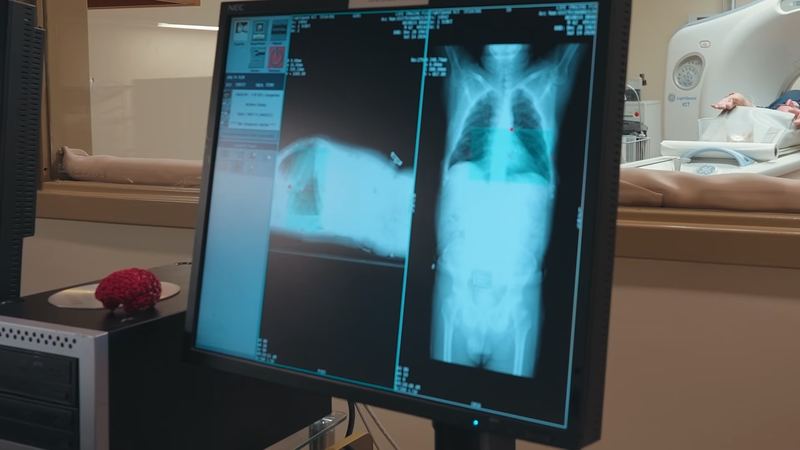
Virtual colonography can detect many polyps, including some that lie flat against the colon lining. Flat polyps can be harder to detect than growths that project clearly into the colon, so the quality of the preparation, the size and shape of the lesion, and the way the images are interpreted all matter.
This does not mean that virtual colonography cannot detect flat polyps. It means no colorectal screening test finds every lesion. Understanding what the scan can show, where its limitations are, and what happens after an abnormal result can help you choose a screening method with realistic expectations.
Quick Answer
Yes, virtual colonography can detect flat polyps. Larger flat lesions and those with a visible surface change may be easier to identify than very small or subtle ones.
Some sessile serrated lesions are particularly difficult to see because they may rise only slightly above the colon lining. Residual stool, fluid, incomplete bowel preparation, or poor colonic expansion can make detection more difficult.
Virtual colonography is used to find suspicious growths, not remove them. If the scan identifies a polyp that requires further evaluation, a traditional colonoscopy may be recommended so the area can be examined directly, removed, or biopsied.
What Is Virtual Colonography?
Virtual colonography, also called CT colonography, uses computed tomography to create detailed images of the colon and rectum. Specialized software allows a radiologist to review the colon from multiple angles and examine both two-dimensional and three-dimensional views.
Unlike a traditional colonoscopy, virtual colonography does not involve passing a long scope through the entire colon. It generally does not require sedation, and most people can return to their usual activities after the appointment.
A short, flexible tube is placed just inside the rectum so the colon can be gently expanded with air or carbon dioxide. The expansion helps separate the walls of the colon and makes the inner surface easier to see on the images.
The National Institute of Diabetes and Digestive and Kidney Diseases explains that virtual colonography can be used to look for polyps, cancer, and other changes in the colon.
What Is a Flat Polyp?
Many people picture a colon polyp as a mushroom-shaped growth attached to the colon wall by a stalk. Some polyps do look that way, but not all of them do.
A flat polyp has little height compared with its width. It may appear as a slightly raised area, a subtle thickening, or a change that spreads along the surface of the colon. Some lesions are completely flat or slightly indented.
The National Cancer Institute explains that colorectal lesions may appear as raised polyps, flat areas, or slightly indented growths. Polyps without a stalk are often described as sessile.
Flat appearance and microscopic type are not the same thing. A flat lesion may be an adenoma, a sessile serrated lesion, or another type of growth. The tissue type usually cannot be confirmed from CT images alone. Removal or biopsy may be needed for a laboratory diagnosis.
Why Can Flat Polyps Be Harder to See?
A polyp that rises clearly into the colon creates a noticeable shape. A flat lesion may blend into the surrounding wall.
Several details can affect whether it stands out.
The Lesion May Have Very Little Height
A broad lesion that rises only slightly may not create an obvious contour. This can make it harder to see than a polyp of similar width that projects farther into the colon.
Residual Stool Can Look Similar
Small pieces of stool can resemble polyps, while stool or fluid covering the colon wall can hide a subtle lesion. Careful preparation and tagging material help the radiologist distinguish remaining bowel contents from tissue.
The Colon Must Be Well Expanded
Collapsed or poorly expanded sections can create folds and crowded surfaces. Adequate expansion helps the radiologist inspect the lining more clearly.
Some Lesions Have Subtle Surface Features
Sessile serrated lesions may be pale, broad, and only slightly raised. These characteristics can make them challenging to detect with image-based screening.
Size Still Matters
Larger polyps are generally easier to identify than very small ones. A tiny, flat lesion may present with less visible surface change and fall below the reporting threshold used for virtual colonography.
Can Virtual Colonography Detect Sessile Serrated Lesions?
It can detect some of them, particularly when they are large enough, and the colon has been well prepared and expanded.
Sessile serrated lesions often develop in the right or upper portion of the colon. They may have a broad base and very little height. Their subtle appearance can make them challenging to detect by both imaging and direct visual examination.
Research available in PubMed has shown that serrated lesions can be detected on CT colonography. One feature that may help is that contrast-tagging material coats the surface of certain lesions, making them more noticeable on images.
However, studies have also found that some flat, high-risk sessile serrated lesions are detected less readily by CT colonography than by traditional colonoscopy. This is an important limitation to discuss when choosing a screening method.
How Does the Radiologist Look for Flat Polyps?
Virtual colonography produces a large set of images rather than a single image. The radiologist reviews the colon carefully using several views.
Three-Dimensional Views
Three-dimensional software creates an image resembling a journey through the colon. This view can help show raised areas and changes in the surface.
Two-Dimensional Views
Cross-sectional images allow the radiologist to examine the colon wall from different directions. They can help show density, confirm whether a finding is attached to the wall, and distinguish a possible lesion from tagged stool.
Supine and Prone Images
Images are often taken with the patient in more than one position. Stool and fluid may move when the patient changes position, while a true polyp usually remains attached to the colon wall.
Contrast Tagging
A tagging agent taken during preparation can mix with remaining stool or fluid. Tagged material appears differently on CT, helping the radiologist separate bowel contents from possible tissue.
Radiologists move between these views rather than relying on one image. This is particularly important when a lesion is broad or only slightly raised.
Why Does Bowel Preparation Matter So Much?
Virtual colonography may not involve sedation, but it still requires bowel preparation. A clean colon gives the radiologist the best chance of seeing the inner surface.
Small amounts of stool or fluid may remain even after preparation. Tagging agents can help identify this material, but preparation instructions still need to be followed carefully.
The process may include:
- A temporary clear-liquid diet
- A laxative or bowel-cleansing medication
- A contrast tagging agent
- Instructions about when to stop eating
- Guidance about regular medications
Your instructions may differ based on the facility and your health history. Do not change diabetes medication, blood thinners, or another prescription without speaking with the appropriate healthcare professional.
If the preparation is incomplete, part of the colon may be difficult to assess. The report may recommend repeating the examination or completing another test.
Does Virtual Colonography Find Every Polyp?
No colorectal screening test finds every polyp.
Virtual colonography performs best at finding larger polyps and masses. Very small, flat, or subtle lesions may be harder to identify. Traditional colonoscopy can also miss lesions, particularly when preparation is poor or a flat polyp blends into the surrounding tissue.
Test performance depends on more than the name of the examination. It is affected by:
- The quality of bowel preparation
- How well the colon is expanded
- The size and shape of the lesion
- Whether stool or fluid obscures the surface
- The imaging equipment and technique
- The experience of the interpreting radiologist
A negative result reduces concern but does not guarantee that no polyp is present.
How Does Virtual Colonography Compare With Traditional Colonoscopy?
Both tests can examine the entire colon, but they do so in different ways.
Virtual colonography creates CT images. It is noninvasive, usually does not require sedation, and generally allows patients to return to normal activities promptly.
Traditional colonoscopy uses a flexible camera to directly examine the inner lining. It usually involves sedation. Its major advantage is that the physician can remove a polyp or take a biopsy during the same procedure.
The NIDDK explains that polyps found during colonoscopy can often be removed and sent to a laboratory for testing.
Virtual colonography is a detection test. If it finds something that needs tissue diagnosis or removal, a traditional colonoscopy is generally the next step.
Which Test Is Better for Flat Polyps?
There is no single answer for every patient.
Traditional colonoscopy allows close visual inspection of the colon lining and immediate removal of suspicious tissue. This can be important when someone has a known history of polyps, inflammatory bowel disease, a hereditary colorectal cancer syndrome, or another high-risk condition.
Virtual colonography may be considered by people who want a noninvasive screening option, prefer to avoid sedation, or cannot complete a traditional colonoscopy. It may also be useful after an incomplete colonoscopy when the physician wants to examine the remaining colon.
If flat or serrated lesions are a particular concern because of your history, discuss that concern before choosing a test. Your physician can help you decide which method is best suited to your risk level.
What Happens If Virtual Colonography Finds a Polyp?
The next step depends mainly on the size and appearance of the finding.
A larger polyp or suspicious mass will generally prompt a referral for a traditional colonoscopy. The gastroenterologist can locate the area, remove the tissue when appropriate, and send it for laboratory examination.
Small findings may be handled differently. Depending on their number, size, and appearance, as well as the patient’s risk factors, the options may include colonoscopy or CT surveillance.
The NIDDK notes that a doctor may recommend a follow-up colonoscopy based on the virtual colonography result. Virtual colonography itself cannot remove the polyp or provide a tissue diagnosis.
Ask who will arrange the follow-up before your scan. Knowing the process in advance can prevent delays if the result needs attention.
Will You Need Another Bowel Preparation?
If a follow-up colonoscopy is needed, a second bowel preparation may be required. Whether both examinations can be completed close together depends on scheduling, the facility, and when the virtual colonography result becomes available.
Some patients prefer to know this before choosing virtual colonography. The scan offers the advantage of no sedation, but an abnormal result may still lead to a sedated procedure.
This does not make the initial test unnecessary. Many people have a negative result and do not need an immediate colonoscopy. It is simply one possible outcome to consider.
Can Virtual Colonography Remove a Flat Polyp?
No. Virtual colonography produces images but does not place an instrument inside the entire colon.
If a flat polyp is found, a gastroenterologist may need to examine it directly during a traditional colonoscopy. Removal can be more technically involved when a lesion is broad or sits close to the colon wall, but many flat polyps can still be removed endoscopically.
A laboratory then examines the tissue to determine the type of polyp and whether it contains precancerous or cancerous cells.
The imaging appearance can suggest that an area is suspicious. It cannot replace tissue analysis when a definite diagnosis is needed.
Are Flat Polyps More Likely to Become Cancer?
Flat shape alone does not determine whether a polyp will become cancer. The risk depends on the tissue type, size, microscopic changes, and other features.
Most colon polyps are not cancer. Some adenomas and sessile serrated lesions can develop into colorectal cancer over time. Larger lesions and those with certain cellular changes may carry greater risk.
Because imaging cannot identify all microscopic features, a removed polyp is usually sent to a laboratory. The pathology result helps determine whether additional treatment or earlier surveillance is needed.
Do not assume that “flat” means cancerous. It describes the lesion’s shape, not its final diagnosis.
Who May Consider Virtual Colonography?
Virtual colonography may be an option for an average-risk adult who is due for colorectal cancer screening and wants an alternative to traditional colonoscopy.
It may also be considered when:
- A previous colonoscopy could not examine the entire colon
- Sedation presents a concern
- A patient prefers a noninvasive examination
- A physician recommends it based on an individual health situation
Virtual colonography may not be the first choice for everyone. Someone with previous advanced polyps, a strong family history, inflammatory bowel disease, hereditary cancer risk, active bleeding, or significant bowel symptoms may need a different evaluation.
A screening test is chosen for a person, not just for a diagnosis. Discuss your history and preferences before deciding.
Can You Have Virtual Colonography If You Have Symptoms?
Virtual colonography may be used in some diagnostic situations, but symptoms should first be discussed with a healthcare provider.
Blood in the stool, unexplained iron-deficiency anemia, ongoing abdominal pain, a persistent change in bowel habits, or unexplained weight loss may require a more direct evaluation. Traditional colonoscopy may be recommended because it allows the physician to inspect the lining and collect tissue during the same procedure.
Do not use routine screening as a substitute for care when symptoms are already present.
What Does a Negative Result Mean?
A negative virtual colonography means the radiologist did not identify a polyp or mass that met the reporting threshold. It is generally reassuring, but it does not guarantee that the colon contains no small or subtle lesions.
Your next screening date will depend on the quality of the examination, your risk level, and the report’s recommendation. Average-risk adults with a complete negative screening examination may be advised to repeat CT colonography after the recommended interval.
If the preparation was incomplete or part of the colon could not be assessed clearly, the result may not be considered fully negative. Another test may be recommended sooner.
Keep a copy of your report and confirm the next step with your healthcare provider.
How Can You Get the Most From Your Examination?
Preparation starts before scan day.
Read the instructions as soon as you receive them. If anything is unclear, ask before beginning the bowel preparation, and waiting until the evening before the test can make it harder to solve a medication or timing question.
Tell the imaging center about:
- Previous colon polyps or colorectal cancer
- A family history of colorectal cancer
- Previous incomplete colonoscopy
- Inflammatory bowel disease
- Current digestive symptoms
- Pregnancy or possible pregnancy
- Prescription medications
- Problems with earlier bowel preparations
Follow the dietary, laxative, and tagging instructions closely. On the day of the examination, tell the technologist if you have severe discomfort or difficulty changing position.
You can learn more about virtual colonography at Life Imaging and review what to expect during your visit.
What Questions Should You Ask Before Choosing a Test?
Your screening choice should be based on more than convenience.
Ask your healthcare provider:
- Am I considered average risk or high risk?
- Is virtual colonography appropriate for my health history?
- Does my history raise concern about flat or serrated lesions?
- What preparation will I need?
- What happens if the scan finds a polyp?
- Will I need a traditional colonoscopy after an abnormal result?
- How often should I repeat screening after a negative result?
- Who will explain the report and arrange follow-up?
There is no need to choose a test based on fear or assumptions. A clear discussion can help you understand what each option can and cannot do.
Frequently Asked Questions About Flat Polyps and Virtual Colonography
Are Flat Polyps Invisible on Virtual Colonography?
No. Virtual colonography can detect flat polyps, especially when they are large enough to create a visible surface change. Very small or subtle flat lesions may be harder to identify than raised polyps.
Can CT Colonography Find Sessile Serrated Polyps?
It can find some sessile serrated lesions. Detection may be more difficult because these lesions can be broad, pale, and only slightly raised. Preparation quality, contrast coating, lesion size, and reader experience can affect visibility.
What Size Polyp Can Virtual Colonography Detect?
Virtual colonography is better at finding larger polyps than very small ones. Findings measuring 6 mm or more commonly receive greater attention in reporting and follow-up decisions. Size is not the only consideration, so the full radiology report should guide the next step.
What Happens If a Flat Polyp Is Found?
A traditional colonoscopy may be recommended so the lesion can be examined directly, removed, or biopsied. The tissue can then be analyzed in a laboratory to determine the polyp type.
Can a Negative Virtual Colonography Miss Cancer?
No screening test provides a guarantee. A complete negative examination is reassuring, but small or subtle lesions can occasionally be missed. Follow the recommended screening interval and report new digestive symptoms rather than waiting for the next routine examination.
Is Virtual Colonography Better Than Traditional Colonoscopy?
Each test has advantages and limitations. Virtual colonography is noninvasive and generally does not require sedation. Traditional colonoscopy allows the physician to view the lining directly and remove polyps during the same procedure. Your risk level, health history, and preferences should guide the choice.
Choose a Colon Screening Plan With Confidence
Virtual colonography can detect many polyps, including some flat and sessile lesions. Its ability to detect specific growths depends on the lesion’s size and appearance, the quality of the bowel preparation, and how clearly the colon can be evaluated. If the scan finds something concerning, traditional colonoscopy may still be needed for removal or biopsy.
Life Imaging provides virtual colonography for patients seeking a noninvasive way to examine the colon. Speak with your healthcare provider about your personal risk, previous polyps, family history, and any current symptoms before choosing a colorectal screening test.